Title: A Comparison of the Anatomies of Sus and Homo sapiens
Background Research:
Sus is a placental mammal, just like humans. The anatomy of the fetal pig resembles that of other placental mammals such as humans. Nearly all major structures are the same or similar in anatomy. For example, it has the same circulatory system, similar muscles, and a similar skeletal structure. Therefore, it allows for a representative overview of vertebrate anatomy and provides the framework for understanding functioning body systems. Fetal pigs are not bred for the purpose of dissection. They are a by-product of the pork food industry. Also, the fetal pigs aren’t ever born, so they do not “die” for dissection purposes. It’s a viable alternative for those concerned about the use of live animals in scientific study. Furthermore, soft fetal tissue is easy to dissect.
The fetal pig’s external physiology can be divided into the following main regions:
- · cranial (head) region
- · cervical (neck) region
- · thoracic region
- · lumbar (trunk) region
- · sacral region
- · caudal (tail) region
Much of the external physiology of the fetal pig is the same as that of humans such as ears, nose, eyes, etc… The fetal pig has four appendages, two forelimbs and two hind limbs. The shoulder, elbow, wrist, and digits are on the forelimb. The hip, knee, ankle, heel, and digits are on the hind limb. On the ventral side of the pig, there is the umbilical cord which connected the pig to its mother’s placenta. If the pig is female it will have a single urogenital opening ventral to the anus. A prominent genital papilla projects from the urogenital opening. If the pig is male it will have a scrotum, a sac-like swelling containing the testes and located ventral to the anus. The male urogenital opening is faintly visible just posterior to the umbilicus. Also, males as well as females have multiple teats or mammary papillae. Also, the fetal pig may have a thin peeling layer of tissue covering its body. This layer is the epitrichium, a layer of embryonic skin that peels off as hair develops beneath it.
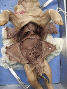
The internal physiology of the fetal pig has 3 cavities which are the oral cavity, the thoracic cavity, and the abdominal cavity.
The oral cavity is the mouth of the pig which contains the papillae (taste buds) on the edges of the tongue, the ridged hard palate in the roof of the mouth with the smooth soft palate behind it, the sharp teeth near the front of the mouth, and the epiglottis, which covers the opening of the trachea (windpipe) so food cannot enter.
The thoracic cavity is protected by the rib cage and contains the following organs:
- · Lungs – the lungs have multiple lobes and are found on either side of the heart.
- · Heart – the heart is encased in a shiny pericardial membrane
- ·.
- · Larynx – at the top of the throat the trachea bulges out into the larynx, or voice box
The abdominal cavity is located below the thoracic cavity and is separated by the diaphragm. It holds the following organs:
- · Liver – the liver is the large black/brown multi-lobed organ at the top of the abdominal cavity.
- · Stomach – the pig’s stomach is located on the right side, tucked under the liver.
- · Spleen – the spleen is not part of the digestive system; it helps filter the pig’s blood. It is a thin finger-like organ lying on the stomach and matching it in color.
- · Small Intestine – the small intestine is a large mass of coiled tube that fills the bottom half of the abdominal cavity. It is held in place by tissue called mesentery. Lift up a section of the intestine and pull it tight – the mesentery is the thin tissue filled with blood vessels.
- · Large Intestine – the large intestine’s big coils look fused together. It sits to the right of the small intestine, just below the stomach.
- · Kidneys – carefully move the intestines aside to see the large bean-shaped kidneys (one on each side) covered in a shiny membrane.
- ·.
Works Cited:
“Fetal Pig Dissection.” Virtual with Pictures. N.p., n.d. Web. 12 Jan. 2013.
“Virtual Fetal Pig Dissection (VPD).” Virtual Fetal Pig Dissection (VPD). N.p., n.d. Web. 12 Jan. 2013.
Data Collection: External
- A. Measurements
R. Fore Limb
20 cm
L. Fore Limb
20 cm
Average
20 cm
R. Hind Limb
20 cm
L. Hind Limb
19 cm
Average
19.5 cm
Circumference
25 cm
Snout
2 cm
Head
12 cm
Torso
29 cm
Tail
7 cm
40 cm
Total Weight
1.4 kg ± 0.2 kg
Uncertainty: ± 0.5 cm (for all length measurements)
- B. Drawings – On separate paper
Data Collection: Internal
- A. Measurements
Length (cm)
Weight (g)
Alimentary Canal
296
17
Uncertainty: ± 0.5 cm and ± 0.5 g
- B. Drawings – on separate paper
Comparison of the anatomies of Sus and Homo sapiens
The overall digestive tract is very similar anatomically in pigs and humans except the pig digestive tract is noticeably shorter than the human digestive tract. Also, pigs do not have an appendix at the end of the caecum like humans do. Instead, in pigs, the caecum has a pouch that holds bacteria that aid digestion. It is an active part of the digestive system. In humans, it has evolved into a vestigial appendix.
The general structure of the Respiratory System in Homo sapiens and Sus is similar. The pig’s respiratory system is significantly smaller in size but both humans and pigs inhale oxygen and exhale carbon dioxide, like other animals. A two-part respiratory system, upper and lower, exists in both. However, in humans, the right lung has three lobes, and the left lung has two lobes. In pigs, the left lung has two lobes, and the right lung has four lobes. Pigs also able to take more breaths in a minute.
Circulatory System
Both Homo sapiens and Sus circulate their blood through a closed circulatory system called the cardiovascular system. Both have a four-chambered heart, seeing as they are both mammals. Both use blood to transport nutrients, wastes, water, oxygen, and carbon dioxide throughout the body. The pig’s circulatory system is smaller in size. Pig has a caudal artery that brings blood to the tail. This is absent in humans.
Urinary System
The urinary system within humans and pigs is very similar in structure and function. It is responsible for processing the waste products of metabolism. The major organs and parts that make up the urinary system in both humans and pigs include the kidneys, ureter, urinary bladder, and urethra. Also, humans and pigs are two examples of how waste removal is a crucial part in maintaining homeostasis. In humans and pigs, the first thing that happens in the urinary system is blood enters the kidneys through the renal artery and exits through the renal vein. As the blood passes through the kidneys, millions of nephrons or excretory tubules within the kidneys filter out blood plasma, nitrogenous wastes, urea, salts, ions, glucose, and amino acids. Then the nephrons reabsorb the substances needed by the body from the filtrate and return them to the blood. Then the waste fluid, urine, leaves the kidneys through the ureter which drains into the urinary bladder where it is temporarily stored. From there, the urine passes through a tube called the urethra and exits the body during urination. The urinary system of a pig and a human is almost identical. The only major difference is the size of the organs between that of a human and that of a pig.
Reproductive System
Female
In female humans and pigs, the main organs that compose their reproductive system consist of the ovaries, oviducts, uterus, and vagina. A difference between female pigs and humans is that a pig’s vagina connects to the urogenital aperture and humans do not have a urogenital aperture. The vagina is a short, muscular canal located below the urethra. Also, the pig’s vagina is much shorter in length than the human vagina. In female pigs, the ovaries which produce egg cells (ova) and female hormones (estrogen and progesterone), are small and located just below the kidneys inside the peritoneal membrane. Also, the shape of the uterus in pigs is different than in humans. In pigs it is Y-shaped and in humans it’s a pear shaped organ. Furthermore, pigs do not undergo menstruation such as humans do. Instead, in pigs, ovulation is provoked by mating.
Male
In male humans and pigs, the major organs that make up their reproductive system include the testes, scrotum, epididymis, prostate glands, seminiferous tubules, vas deferens, seminal vesicles, urethra, and the penis. In both humans and pigs, the testes located in the scrotum, are the reproductive organs that produce sperm and male hormones (testosterone). The seminiferous tubules which are tightly packed coils located in the testes are the place in which sperm develop. In pigs and humans, sperm are stored in the epididymis which is adjacent to the testes. Also, pigs and humans both produce seminal fluid inside the prostate gland and seminal vesicles. A difference between pigs and humans is that pigs have a urogenital aperture and humans do not. Another major difference between pig and human male reproductive systems is that, during the non-breeding periods, pigs possess the ability to retract the testes into the abdominal cavity. Humans cannot do this.
Works Cited
BSCS Biology: A Molecular Approach. Columbus, OH: Glencoe/McGraw-Hill Publishing Company, 2006. Print.
Bohensky, Fred. Photo Manual and Dissection Guide of the Pig. USA: Square One Education Guides, 2002. Print.
Kimball, John. Kimball’s Biology Pages. 2010. Web. 2 June 2010.
National Kidney and Urological Diseases Information Clearinghouse. n.d. Web. 2 June 2010.
Pig Breeding Giode. n.d. Web. 2 June 2010.
Damon, Alan. Biology: Higher Level (plus Standard Level Options) : Developed Specifically for the Ib Diploma . Harlow: Heinemann, 2008. Print.