Sadie, a five-year-old Golden Retriever, clearly wasn’t her normal self. Her adoptive human mom, Leah, wished the dog could tell her if she hurt or what was making her so restless. The next time she approached Sadie, she noticed that the dog’s right eye appeared to be drooping. A visit to the veterinarian the same day confirmed Horner’s syndrome in dogs.
What is Horner’s Syndrome?
This condition is really a group of signs that appear when certain facial muscles in a dog are no longer stimulated by the sympathetic nerves, according to PetEducation.com. While the syndrome can affect any breed of dogs, for reasons experts haven’t been able to identify, it appears to be the most common in Golden Retrievers.
In up to half the dogs affected, vets never find the cause of the disorder. They classify this type of Horner’s syndrome as idiopathic. However, they know that in some dogs, a brain injury or brain stem lesion can cause the problem, PetMD reports. Other causes that have been identified are trauma from car accidents, disc disease in the neck area, bite wounds, middle-ear infections, disease in the orbit of the eye, cancer and treatment such as cleaning or administering medication.
When Horner’s syndrome results from an injury, the location might occur at the level of the dog’s brain, upper spinal cord or between the face and spinal cord. The dog’s symptoms will appear on the same side of the head as the location of the injury.
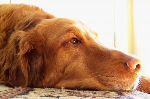
There are several signs of Horner’s syndrome in dogs. This nerve disorder can produce a drooping eye, a severely constricted pupil in the dog’s eye or a protruding eyelid. An exam might show a smaller pupil (known as miosis) in the affected eye than in the normal one.
Many owners notice an abnormal elevation of their dog’s inner, or third, eyelid. Sometimes the dog’s eye appears sunken into its socket. Ear inflammation isn’t uncommon.
Diagnosis and Treatment
Horner’s syndrome is relatively easy for vets to diagnose based on symptoms alone. However, what is far more difficult and sometimes impossible is finding out what caused it to appear in the dog.
In addition to a standard health history and asking the owner about symptoms and dates they appeared, a vet should perform a physical examination that includes a neurologic evaluation. Most practitioners order X-rays, a blood chemistry panel, urinalysis and a complete blood count (CBC). In some cases, CT, MRI or ultrasound scans are necessary to help determine what caused the disorder. A cerebrospinal fluid (CSF) sample can indicate brain and spinal cord disease.
In checking for the site of any eye injury, vets typically administer epinephrine to the dog’s eye and note the amount of time it takes the pupil to dilate. The elapsed time helps pinpoint where along the pathway of the brain and spinal cord the injury occurred.
There is no treatment for Horner’s syndrome itself. However, vets need to treat the underlying cause if they can find it. If, for example, there is an ear bite, it would need to be treated, along with any infection that had developed. A dog with a middle-ear infection would receive standard treatment for that condition.
Depending on the location of the problem, phenylephrine eye drops can relieve clinical signs associated with the disorder. When the cause of Horner’s syndrome is idiopathic, the condition often resolves on its own in six to eight weeks.
Sources:
http://www.peteducation.com/article.cfm?c=2+2105&aid;=2478
http://www.petmd.com/dog/conditions/eyes/c_dg_horners_syndrome


